System: Skin: Vascular: Benign: Cavernous Hemangioma


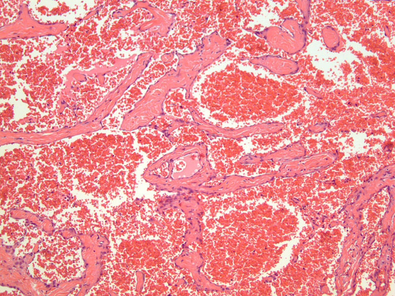
Case 1: Blood filled vascular spaces lined by attenuated endothelial cells is characteristic. The walls are often fibrotic.
Similar to capillary hemangiomas, cavernous hemangiomas tend to affect children. However, unlike capillary hemangiomas, they are more likely to be larger, deeper and are less likely to regress spontaneously. In additional to cutaneous locations, cavernous hemangiomas are the most common intraorbital tumors in adults, and have also be described in the liver and heart.
Grossly, they are irregular, red lesions when they are on the superficial skin. The cut surface is spongy. Histologically, they are composed of dilated, blood-filled spaces lined by flattened endothelial cells. The walls are often fibrotic. Thrombosis and dystrophic calcifications are common findings (Folpe).
They may be associated with Kasabach-Merritt syndrome (consumption coagulopathy) and Marfucci syndrome (hemangiomas, multiple enchondromas)(Fletcher, Folpe).
→Tends to be larger, deeper and less likely to regress compared to capillary hemangiomas. Most common intraorbital tumor in adults. Can arise in the liver and heart.
→Association with Marfucci syndrome (hemangiomas and enchondromas), Kasabach-Merritt syndrome (consumption coagulopathy).
• Vascular : Spindle Cell Hemangioma
Fletcher CDM, ed. Diagnostic Histopathology of Tumors. 3rd Ed. Philadelphia, PA: Elsevier; 2007: 48.
Folpe AL, Inwards CY. Bone and Soft Tissue Pathology: Foundations in Diagnostic Pathology Philadelphia, PA: Elsevier; 2010: 172-3.
)