
Islands of odontogenic epithelium are embedded in a loose stroma. Note the presence of amorphous material that may be enamel or dentin.
)
Thin cords of odontogenic epithelium embedded in a primitive ectomesenchyme; this area is indistinguishable from ameloblastic fibroma.
)
Another image with the components of odontogentic epithelium, mesechyme and dentin or enamel material.
)
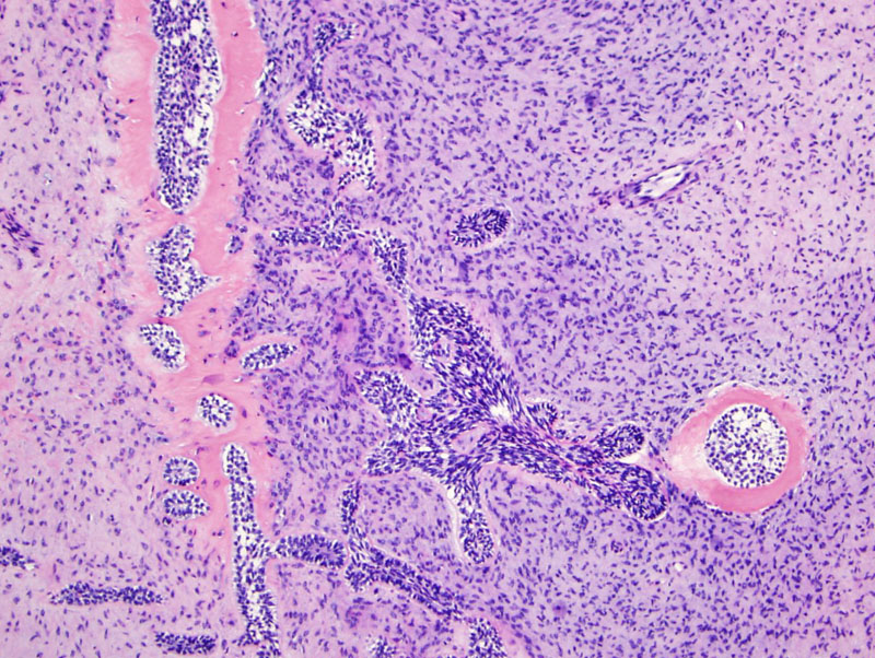
The calcified material approximates the odontogenic epithelium.
)
The mesenchymal component is likened to dental papillae of developing teeth.
)
The odontogentic epithelium has two layers, an outer layer with darker basaloid cells that resemble ameloblasts and an inner loose layer that resembles stellate reticulum.
)
This second case also consists of anastamosing cords of odontogenic epithelium in a loose stroma.
)
The odontogenic epithelium can form islands, cords or strands.
)
Note the presence of dentin or enamel matrix next to the odontogentic epithelium.
)
Short cords of odontogenic epithelium within a loose myxoid stroma. Both components are bland, without atypia or hypercellularity.
)
Ameloblastic fibroma (AF) and ameloblastic fibro-odontoma (AFO) are related neoplasms composed of odontogenic epithelium and mesenchyme; fibro-odontomas have the added components of dentin and enamel.
Histologically, ameloblastic fibromas are composed of cords, strands and islands of odontogenic epithelium embedded in a loose stroma that resembles embryonic dental pulp. The palisading peripheral cells in the epithelial islands are basaloid and resemble ameloblasts -- whereas the central cells are looser and resemble reticulum-like tissue. In ameloblastic fibro-odontomas, mineralized material (enamel, dentin) is also present in the stroma (Barnes, Thompson).
These are uncommon tumors that occur within the first two decades of life (mean age between 8-12). AFOs are often asymptomatic, and often associated with an unerupted tooth. Ameloblastic fibromas have a slight male predilection whereas ameloblastic fibro-odontomas have equal gender distribution. The posterior mandible is the most common location.
Radiographically, there is a well-circumscribed unilocular or multilocular lucent area with areas of opacity (depending on the extent of mineralization).
A subset of ameloblastic fibromas do occur and uncommonly, may transform into ameloblastic fibrosarcomas. Ameloblastic fibro-odontomas, however, are not known to recur.
→Ameloblastic fibroma, ameloblastic fibrodentinoma and ameloblastic fibro-odontomas are related entities composed of amelolastic epithelium embedded in a loose pulp-like stroma.
→These are rare entities that arising mostly in young individuals, between age 8-12.
• Oral Cavity : Ameloblastic Fibroma
Barnes L, Eveson JW, Reichart P, Sidransky D. WHO Classification: Pathology and Genetics, Head and Neck Tumors Lyons, France: IARC; 2005: 308-9.
Thompson LDR, Wenig BM, eds. Diagnosis Pathology: Head and Neck. 1st Ed. Manitoba, Canada; Amirsys;2011; 6-48,49.