System: Head and Neck: Sinonasal: Neoplastic: OVERRIDE

System: Head and Neck: Sinonasal: Neoplastic: OVERRIDE

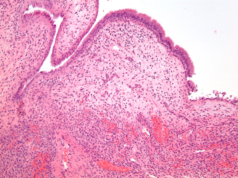
Case 2: Normal intact sinonasal respiratory epithelium overlies the tumor.
The spindle cells have bland oval nuclei and are arranged in a vague fascicles.
Diffuse SMA staining supports the myoid origin of these cells.
Case 3: Another case with a monotonous proliferation of ovoid cells. Extravasated RBCs are a common finding.
Scattered inflammatory cells are common. Note that in contrast to solitary fibrous tumors, collagen fibers are not a prominent feature.
The vessels may be staghorn-shaped or simply irregular.
Perivascular hyalinization is a helpful finding seen in the majority of glomangiopericytomas, however, in this case, the hyalinization is a bit subtle.
)
)
)
)
)
)
)