System: Skin: Melanocytic: Neoplastic: Melanoma, Balloon Cell Type

System: Skin: Melanocytic: Neoplastic: Melanoma, Balloon Cell Type

low power shows effacement of dermal architecture by sheets of neoplastic cells with no intervening stroma nodular proliferation of neoplastic balloon cells, typically the background is one of a superficial spreading melanoma Image
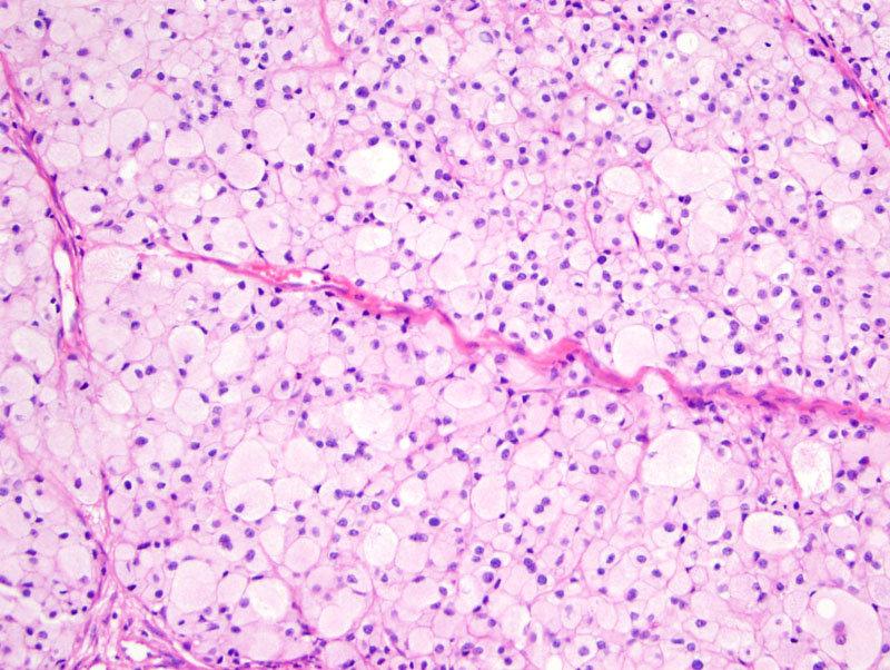
sheets of large cells with abundant quantity of clear or finely vacuolated cytoplasm; Image
clearness of cytoplasm can be due to intracellular accumulation of glycogen ; have to differentiate from balloon cell nevus Image
studies with antibodies to S100 protein, Melan-A and HMB-45 are positive in balloon cell melanoma and its metastases
differential diagnosis encompasses balloon cell change in benign nevi as well as other malignant clear cell neoplasms including clear cell sarcoma of soft parts, atypical fibroxanthoma and granular cell carcinoma with clear cell change, metastatic renal cell carcinoma, clear cell basal cell carcinoma, and malignant clear cell acrospiroma115, sebaceous carcinoma and clear cell squamous cell carcinoma.
soft, rubbery, or firm nodules with a polypoid or papillomatous contour
prognosis is similar to that of other types of melanoma matched for depth of invasion, with tumor thickness being of greatest importance
Magro CM, A Neil Crowson AN, and Martin C Mihm MC, Jr. Modern Pathology (2006) 19, S41–S70. Unusual variants of malignant melanoma.
)
)
)