System: Breast: Ductal: Pre-Neoplastic: Ductal Carcinoma In Situ, Apocrine Type

System: Breast: Ductal: Pre-Neoplastic: Ductal Carcinoma In Situ, Apocrine Type

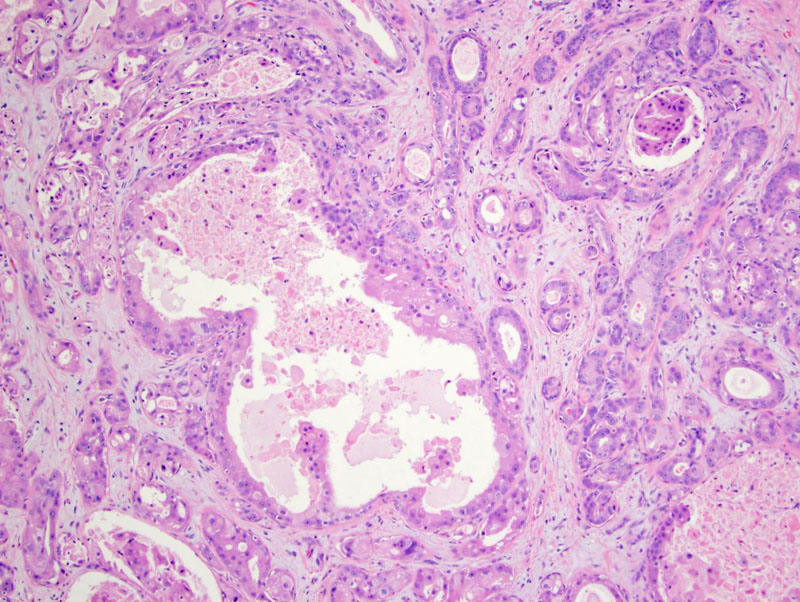
this example of apocrine DCIS is one of that involving extensive sclerosing adenosis, making it look like invasive ca Image
irregular ductal structures are those of adenosis, lined by cells with lots of pink cytoplasm Image
here is a nice area Image
see the apocrine cells arranged in an architecture of dcis Image
larger and smaller ducts, can see a two cell layer if you look carefully Image
cribriform areas Image
smooth muscle actin confirms that it is indeed an in situ process even though so complex looking Image
you can see the apocrine cells in this duct, surround by p63 positive basal cells Image
)
)
)
)
)
)
)
)