System: Genitourinary: Kidney: Neoplastic: Epithelioid Angiomyolipoma

System: Genitourinary: Kidney: Neoplastic: Epithelioid Angiomyolipoma

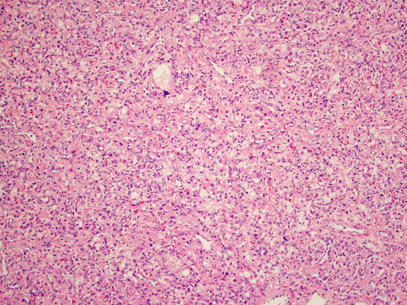
low power, moderately cellular, haphazard solid growth in this field, diagonsis not immediately apparent Image
actually some pleomorphism and some hyperchromasia Image
other aresa with vessels with prominent eosiophilic thickened walls and mature fat Image
cellular areas merge with these funny thick walled vessels of various sizes/calibre Image
another area with the myoid component which predominantes, gives it the "epithleioid"look Image
vessel amongst the myxoid stuff Image
embedded within the fatty areas are these vessels with prominent walls Image
the myoid area and SMA immunostain Image
Pax 8 Immunoreactivity for HMB45, HMB50, CD117, CD63, and negativity for epithelial markers and cytokeratins confirm the diagnosis Image
Clonal
Originate from PEComa cell?
accumulating evidence that this is a malignant neoplasm
associated with tuberous sclerosis
Unlike typical angiomyolipoma for which resection alone is adequate, resection perhaps adjuvant therapy should be considered for this tumor.
Unlike classic renal angiomyolipoma, commonly benign, renal epithelioid angiomyolipoma can behave in an aggressive manner.
- need to recognize so as to not misdiagnose as renal cell carcinoma
)
)
)
)
)
)
)
)
)