System: Genitourinary: Kidney: Neoplastic: Renal Cell Carcinoma, t(6;11) Associated

System: Genitourinary: Kidney: Neoplastic: Renal Cell Carcinoma, t(6;11) Associated

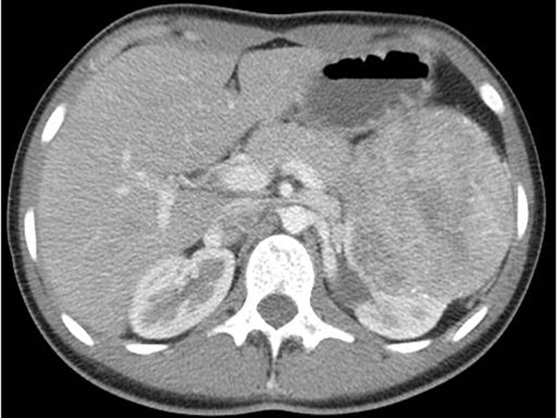
CT scan Image
cut section Image
tumor normal interface Image
1 Image
2 Image
3 Image
Suspect when variable morphology, difficult to classify Biphasic: clear cell /epithelioid cells with small cell secondary population around hyaline BM deposits Image
5 Image
6 Image
7 Image
8 Image
9 Image
ihc Image
pax2 Image
TFE3 (left) and TFEB (right) Image
this tumor on left, epithelioid angiomyolipoma on right Image
t(6;11) TFEB-ALPHA
Rare (~20 reported cases)
Epidemiology not well characterized – may be similar to Xp11 cases
Most cases low stage at diagnosis, without recurrence following resection
)
)
)
)
)
)
)
)
)
)
)
)
)
)
)
)