System: Gynecological: Placenta: Developmental: Diffuse Chorioamniotic Hemosiderosis

System: Gynecological: Placenta: Developmental: Diffuse Chorioamniotic Hemosiderosis

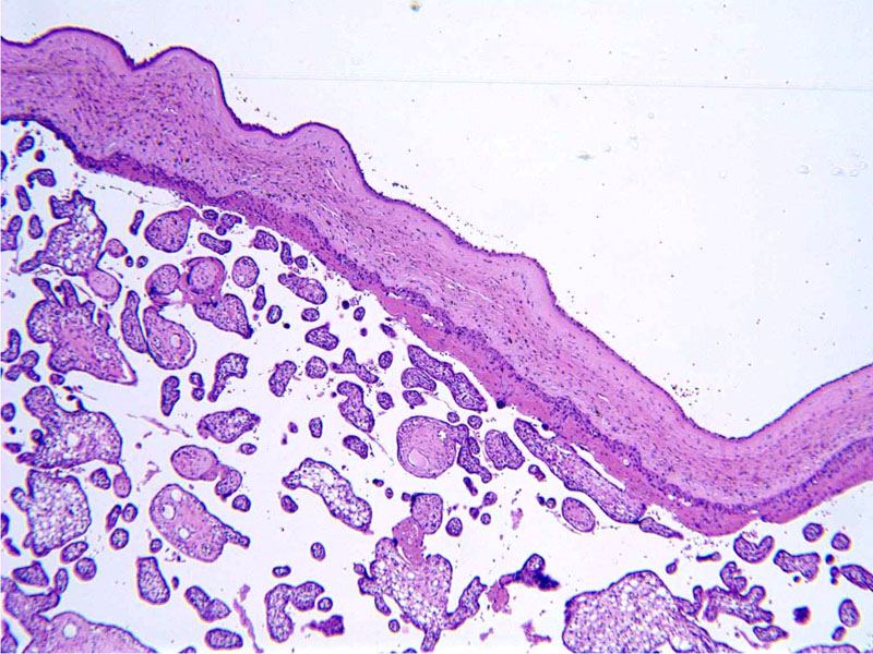
low power of felta side Image
chorion and amnion with brown pigment Image
Extensive hemosiderin deposits in the chorioamnionic membranes Image
pigment Image
areas with iron stain Image
lots of blue Image
gross, maybe put this image first Image
Usually seen in the context of chronic peripheral separation of the placenta or marginal abruption.
Placentas with DCAH more likely to show circumvallation, old peripheral blood clots, increased chorionic villous macrophages, and green discoloration
Associated clinical factors include:
multiparity, smoking and chronic vaginal bleeding were significantly associated
IUGR and oligohydramnios increased but not significantly
Gestational hypertension and advanced maternal age were significantly decreased
No apparent risk of development of neurologic impairment (Redline)
Redline R, Wilson-Costello D. Am J Clin Pathol 1999;111:804
)
)
)
)
)
)
)