System: Gynecological: Placenta: Developmental: Laminar Necrosis Decidual Capsularis

System: Gynecological: Placenta: Developmental: Laminar Necrosis Decidual Capsularis

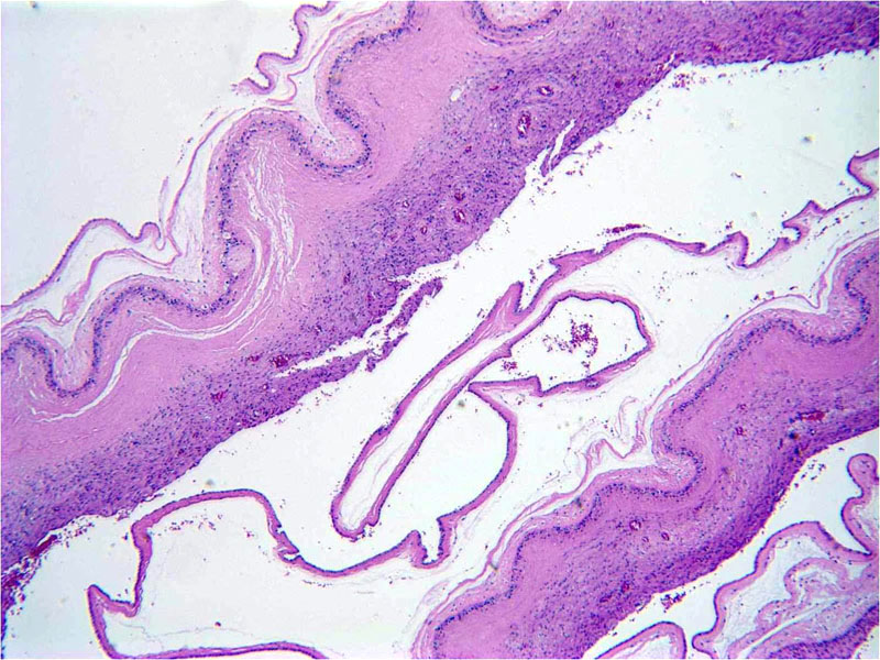
Band-like distribution of coagulative necrosis at the choriodecidual interface of the free membranes. It is also called bland necrosis or leukocytoclastic necrosis (latter term used when karyorrhectic debris is present in addition to ghost outlines of decidual cells) Image
Neutrophils can be seen on the decidual side, otherwise necrosis is non-inflammatory It must be distinguished from patchy decidual necrosis particularly that seen with acute chorioamnionitis Image
3 Image
LN can also involve the basal decidua and has different clinical associations than when confined to the extra-placental (free) membranes.; Here one can see a band-like distribution of coagulative necrosis admixed with a deeper region containing karyorrhectic debris, and it involves at least 30% or more of maternal surface in one slide. LN-decidua basalisis is described as being more common in preterm placentas (23-32 weeks EGA). This type (DB) is associated with pre-eclampsia, indicated preterm birth and lower birth weight and inversely associated with maternal and newborn inflammatory conditions. Appears to be a marker of vascular compromise in preterm infants Occurs more frequently with LN of the membranes Image
Usually involves the decidual layer and frequently the chorionic trophoblastic layer
Involves 10-80% of a membrane roll
Preferably diagnosed when at least 25% of membrane roll involved
Seen in 2nd and 3rd trimester placentas
This under-reported entity is associated with maternal hypertensive disorders, and also with other clinical or placental abnormalities regarded as indicators of uteroplacental hypoxia
)
)
)
)