System: Soft Tissue: Uncertain Lineage: Neoplastic: Ewing's Sarcoma

System: Soft Tissue: Uncertain Lineage: Neoplastic: Ewing's Sarcoma

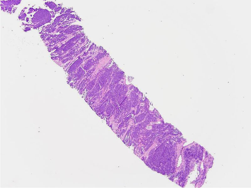
core Image
pre op cytology Image
large intra-abdominal mass Image
maybe we don't need this OR pic, it is interesting though Image
Excised specimen Image
Gelatinous cut surface, looks highly vascular too, encapsulated Image
I am not really sure what this is exactly Image
hi power of the texture of the tumor gross, fenestrated or fibrous septations delicately divide tumor Image
touch prep Image
low power shows vascular areas Image
its in a vessel Image
anothe rnice cytology Image
IHC chart Image
CD99 Image
we put the FISH in the other case so I put this in Image
)
)
)
)
)
)
)
)
)
)
)
)
)
)
)