System: Soft Tissue: Myxoid: Neoplastic: Nerve Sheath Myxoma

System: Soft Tissue: Myxoid: Neoplastic: Nerve Sheath Myxoma

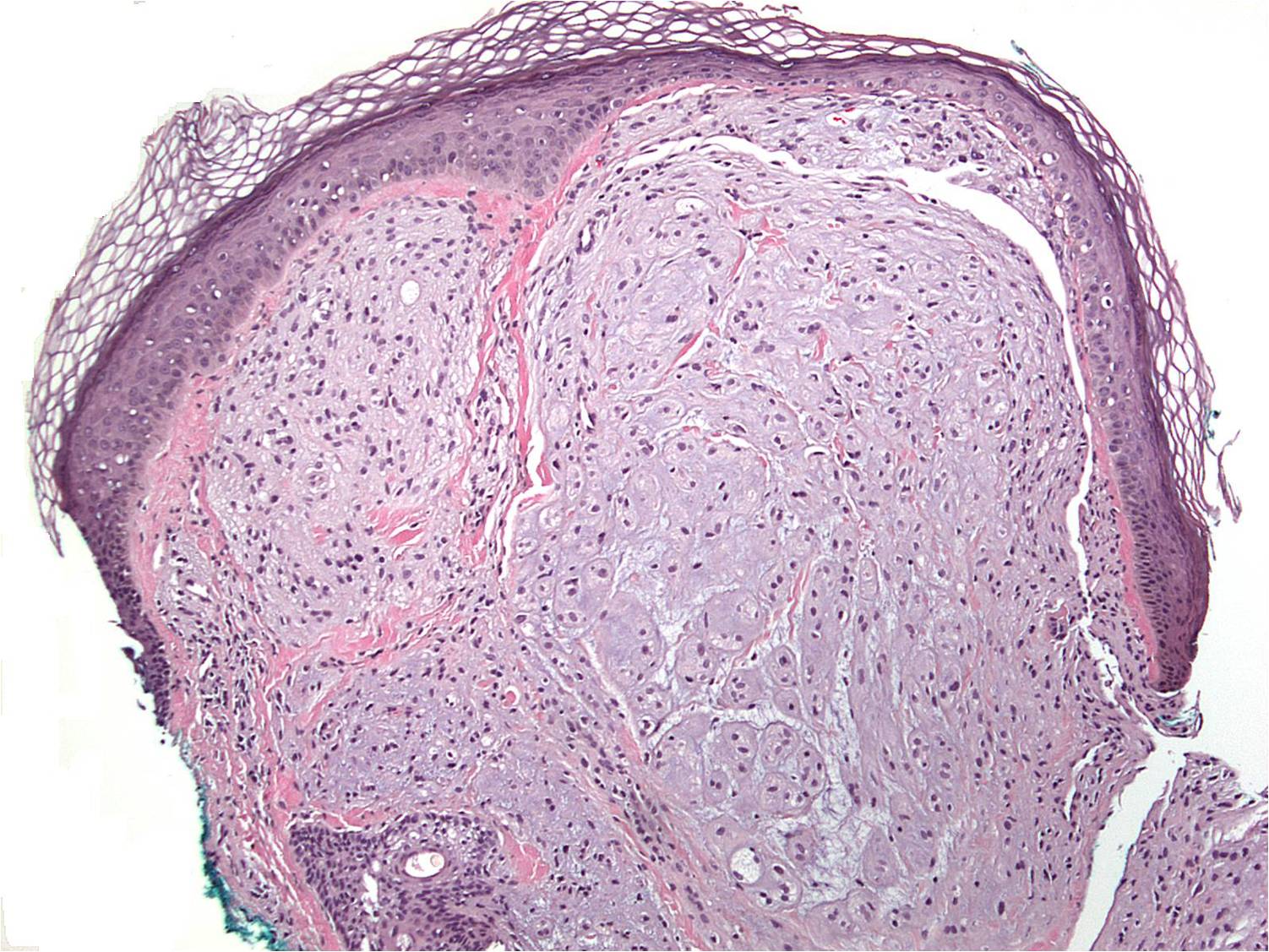
Case 1: The tumor is composed of multiple nodules of cells embedded in a myxoid matrix.
The stellate cells within the nodules are neoplastic Schwann cells, which may form cords or knot-like aggregates.
The differential diagnosis includes myxoid NTK and other myxoid tumors such as myxofibrosarcoma.
Here are the two entities: nerve sheath tumor and myxofibrosarcoma for comparision.
Dermal nerve sheath myxomas used to be considered synonymous with the mxyoid variant pf neurothekeomas used to be considered synonymous with , however, many experts believe this is no longer the case. Myxoid NTKs and true dermal nerve sheath tumor have been demonstrated to be distinct separate entities (Fetsch, 2005).
In an analysis of 178 NTKs (Fetsch et al 2007), the median age of diagnosis was 17. The typical presentation was that of a solitary, superficial nodule with the majority arising in the head, upper extremities and shoulder girdle. In contrast, nerve sheath mxyomas tend to arise predominantly in the extremities, with a median age of 34 years Furthermore, in contrast to NTKs which rarely recurrence, nerve sheath myxomas have a high local recurrence rate.
Histologically, neurothekeomas were composed of multiple small or medium sized nodules separated by collagen bands. The nodules had a whorled pattern and contain variable amounts of myxoid matrix. Osteoclast-like giants were common. The tumor cells were positive for vimentin, NKI/C3, CD10, MTF and PGP9.5, and were negative for S-100, GFAP and MelanA (Fetsh 2007).
Nerve sheath tumors are also composed of multinodular masses with myxoid matrix, however, the nodules contained Schwann cells in cords, nests and syncytial knot-like aggregates. This feature is not seen in neurothekeomas. Furthermore, the neoplastic (Schwann) cells in nerve sheath tumors are positive for S100, GFAP, NSE and CD57 (Fetsh, 2005).
• Myxoid : Myxofibrosarcoma, Low Grade
Hornick JL, Fletcher CD. Cellular neurothekeoma: detailed characterization in a series of 133 cases. Am J Surg Pathol. 2007 Mar;31(3):329-40.
Fetsch JF, Laskin WB, Hallman JR, Lupton GP, Miettinen M. Neurothekeoma: an analysis of 178 tumors with detailed immunohistochemical data and long-term patient follow-up information. Am J Surg Pathol. 2007 Jul;31(7):1103-14.
Fetsch JF, Laskin WB, Miettinen M. Nerve sheath myxoma: a clinicopathologic and immunohistochemical analysis of 57 morphologically distinctive, S-100 protein- and GFAP-positive, myxoid peripheral nerve sheath tumors with a predilection for the extremities and a high local recurrence rate. Am J Surg Pathol. 2005 Dec;29(12):1615-24.
)
)
)
)