
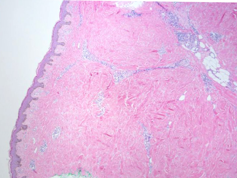
Morphea is characterized a hyalinized collagenous dermis and a perivascular lymphocytic infiltrate. The infiltrate can extend down into the subcutaneous fat. The epidermis can be atrophic or normal. Due to the thick indurated plaques of morphea, the biopsy is often squared off or so-called box-shaped.
)
Note the collagenous hyalinzed dermis with a sparse perivascular lymphocyte infiltrate. The collagen fibers have decreased clefts (very dense) and not many cells (dermal sclerosis).
)
The lymphoplasmacytic infiltrate extends down to the subcutaneous fat, not uncommon in morphea.
)
Morphea is a form of localized scleroderma. It is characterized by similar skin lesions as seen in scleroderma (thickening of dermis and subcutaneous tissues), however, unlike scleroderma, there is no systemic involvement. There is some evidence that a subset of cases (less than 5%) may progress to scleroderma (Nyugen).
Indurated plaques on the skin is seen on skin clinically, especially on the trunk.
Nguyen JV. Morphea: eMedicine. Last updated on Jan 29th 2010. Available at: emedicine.medscape.com/article/1065782-overview
Rapini RP. Practical Dermatopathology. Philadelphia, PA: Elsevier; 2005: 131-2.