System: Gastrointestinal: Small Intestines: Developmental: Microvillous Inclusion Disease

System: Gastrointestinal: Small Intestines: Developmental: Microvillous Inclusion Disease

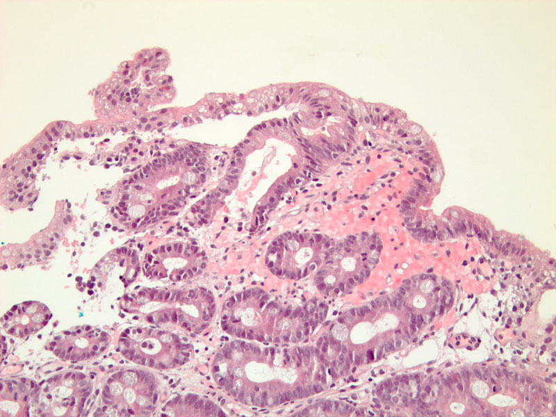
altered architecture with marked villous atrophy Image
granules accumulating in the apical cytoplasm of mature enterocytes and an altered enterocyte brush border membrane Image
CD10 reveals ean nlarged intracytoplasmic band along the apical pole of enterocytes (corresponding to autophagocytic vacuoles and microvillous inclusion bodies revealed by EM)
Presents in the few days of life with severe secretory diarrhea[ metabolic acidosis and dehydration rapidly sets in
Many infants do not survive the first three years of life, and sucumb to infectious complications or liver failure.
)
)
)