System: Soft Tissue: Uncertain Lineage: Neoplastic: Epithelioid Sarcoma, Proximal Type

System: Soft Tissue: Uncertain Lineage: Neoplastic: Epithelioid Sarcoma, Proximal Type

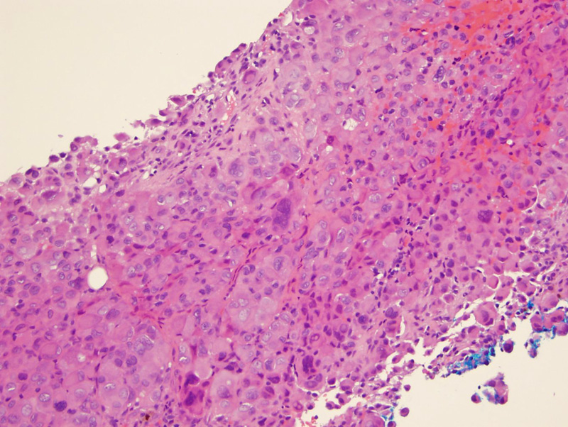
sheets of large cells with prominent nucleoli resembling poorly differentiated carcinoma Image
this tumor resembles other epithelioid lesions, such as extrarenal rhabdoid tumor, epithelioid angiosarcoma and melanoma; has a rhaboid appearance Image
3 Image
4 Image
IN1 1 loss is characteristic of both regular and proximal type epithelioid sarcoma (Hornick). The inflammatory cells in the background show normal nuclear staining. Image
EMA --- positive in about 85% of cases. while many sarcomas can have cytoplasmic EMA, this tumor along with synovial sarcoma, show nice membrane postivitiy. Focal cytoplasmic staining for muscle markers, either desmin or alpha-smooth muscle actin (SMA), S-100 protein, neurofilament, neuron-specific enolase, synaptophysin, and CD56 may be found in a minority of cases. Image
CAM 5.2; this is one of the tumors that co-expresses both vimentin and keratin Image
predominantly in the deep soft-tissue or subcutis of young adults or middle-aged individuals and had a significant male predominance
The rate of tumor-related deaths is lower than the rate of metastasis (75%), suggesting an indolent yet persistent nature for this tumor. Important adverse prognostic factors are larger tumor sizeand early metastasis, the latter independently correlating with clinical aggressiveness.
Hornick JL, Dal Cin P, Fletcher CD. Loss of INI1 expression is characteristic of both conventional and proximal-type epithelioid sarcoma. Am J Surg Pathol. 2009 Apr;33(4):542-50.
)
)
)
)
)
)
)