
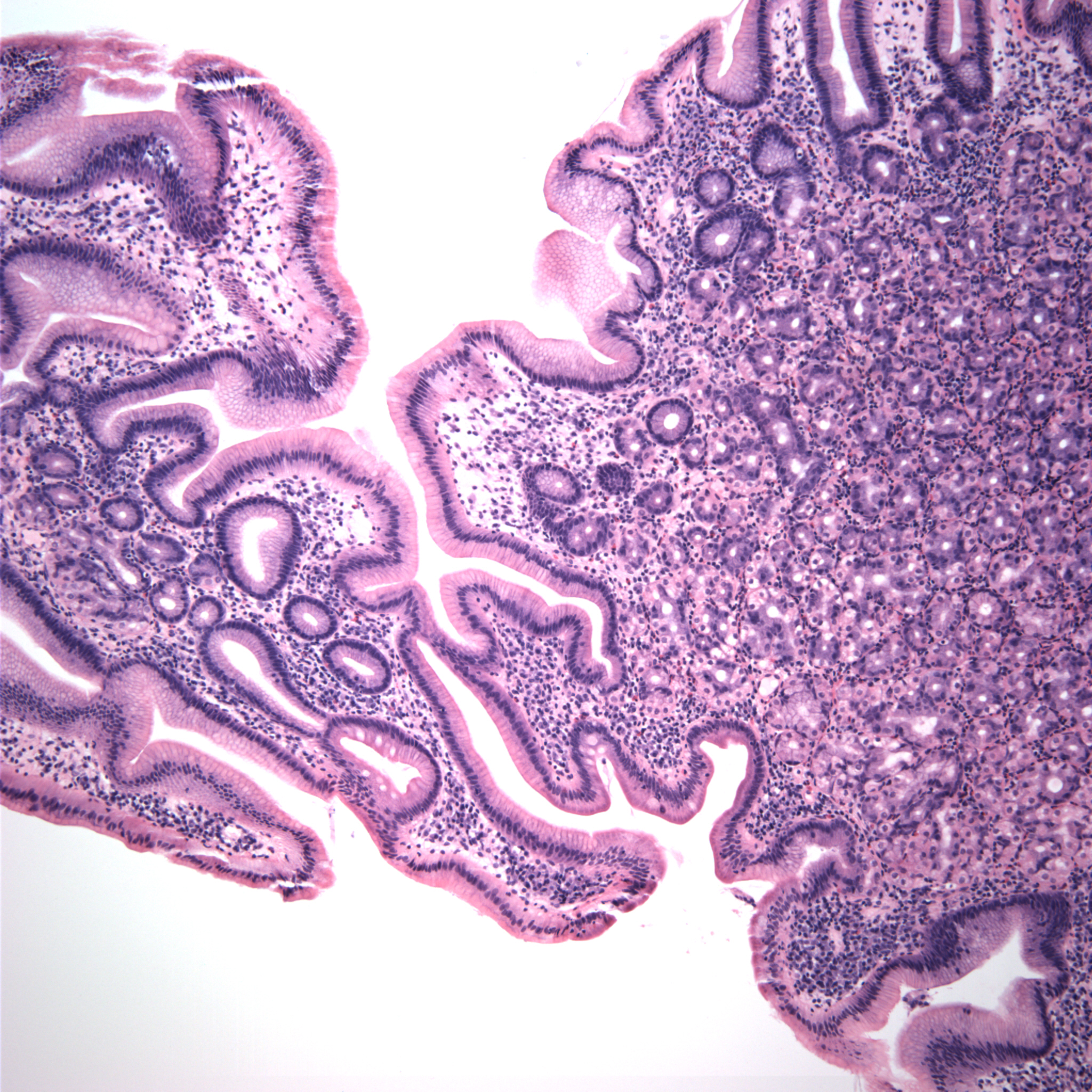
Case 1: Biopsy of a nodule at the duodenal bulb. Duodenal villi can be seen on the left side of image and a heterotopic rest of gastric glands can be seen on the right, underneath foveolar-type mucinous epithelium.
)
A closer look reveals the pink parietal cells and the more basophilic chief cells.
)
On the same biopsy, normal duodenum with Brunner glands can be appreciated.
)
Gastric heterotopia in the duodenal is most commonly identified in the bulb. Endoscopically, these heteropic rests are seen as small (<1.0cm) polypoid nodules. Microscopically, there are normal gastric glands composed of chief and parietal cells. The intestinal epithelium of the overlying surface are often replaced by gastric foveolar-type mucinous epithelium with adjacent normal duodenal mucosa.
Usually benign without clinical significance unless large enough to cause obstruction.
• Small Intestines : Heterotopic Pancreas
Contributed by Dr. Kate Sciandra, Dept of Pathology, VAMC Albuquerque New Mexico.