
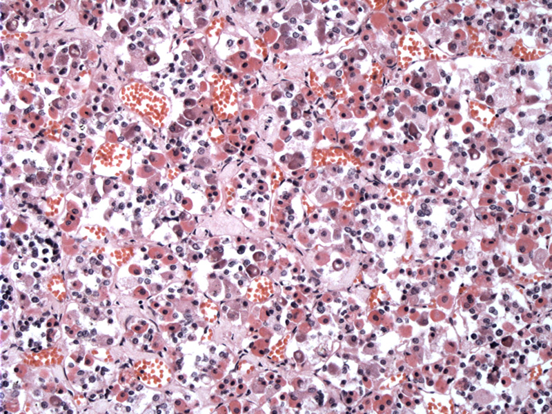
Packets of chromophobes and chromophils comprise the pars distalis. A reticulin network runs between the clusters of cells, but is not visible unless a reticulin stain is performed.
)
At higher power, the two types of chromophils can be distinguished clearly. The acidophils are pink and basophilis are purple. The chromophobes have clear cytoplasm, but due to fixation, their outlines are not clearly delineated here.
)
Here is my rendition of the pituitary gland. I confess I am not an artist, but hopefully, the drawing demonstrates that the gland is composed of the anterior and posterior portions. The posterior pituitary (neurohypophysis) is an extension of the hypothalamus.
)
The pituitary gland, also known as the hypophysis, is composed of two structures with different embryological origin. The anterior pituitary forms from an outpouching of the epithelium from the oral cavity "Rathke's pouch" while the posterior pituitary forms as an extension of the developing brain. The two intertwine to form the pituitary gland.
The anterior pituitary is composed of three subdivisions: pars distalis (the largest subdivision and contains cells which elaborate the important hormones), pars intermedia (a thin layer that separates the pars distalis from the posterior pituitary) and pars tuberalis (surrounds the infundibulum).
The cell types found within the pars distalis are either chromophils or chromophobes. Chromophobes (like their name) are pale-staining and thought to be either degranulated chromophils or stem cells. Chromophils are further subdivided into acidophils (somatotrophs, mammotrophs) or basophils (gonadotrophs, thyrotrophs and corticotrophs). Somatotrophs secrete somatotrophic, mammmotrophs secrete prolactin, thryotrophs secrete thyroid-stimulation hormone (thyrotropin), gonadotrophs secrete FSH and LH and corticotrophs secrete ACTH.
The posterior pituitary also is composed of three subdivision: pars nervosa (the largest subdivsion and contains axons of cells whose cell bodies are located in the hypothalamus. Nuclei seen in this division belong to supportive cells called pituicytes), infundibulum (stalk) and the tuber cinerum of the median eminence.
Eroschenko VP. diFiore's Altas of Histology with Functional Correlations. 10th Ed. Philadelphia, PA: Lippincott Williams & Wilkins; 2005: 329-335.