System: Hematopathology: Spleen: Benign: Littoral Cell Angioma

System: Hematopathology: Spleen: Benign: Littoral Cell Angioma

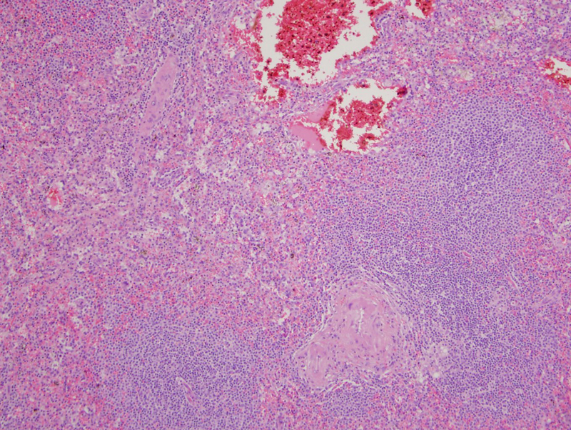
irregular border betwen uninvolved spleen (lower) and the angioma Image
various sized spaces lined by cells with clear cytoplasm Image
blood in cystic spaces Image
another area Image
images from other case Image
s Image
2 Image
cd31 Image
cd34 only vessels Image
lysozyme Image
The most common primary tumors of the spleen are vascular tumors which range from benign lesions (e.g. hemangiomas, lymphangiomas) to more malignant entities (e.g. hemangioendothelioma, angiosarcoma). Littoral cell angioma of the spleen are unique in that it exhibits both vascular and histiocytic markers.
Littoral cells are the reticuloendothelial cells lining the splenic sinuses that demonstrate dual differentiation (endothelial and histiocytic). Thus, they will be immunoreactive for endothelial markers (CD31 and factor VIII) and histiocytic markers (lyzozyme, CD68). Of note, CD34 highlights the vessels only and do not stain the tumor cells (Rosai).
Histologically, contorted vascular channels are lined by plump bland tumor cells. Occasionally, eosinophilic globules may be present in the cytoplasm. The vascular channels can be quite dilated and form "blood lakes". Sloughed tumor cells and pseudopapillary structures may be seen within the dilated spaces.
excision is curative
benign but associated with other visceral malignancies, interestingly
)
)
)
)
)
)
)
)
)
)