System: Skin: Adnexal: Neoplastic: Porocarcinoma

System: Skin: Adnexal: Neoplastic: Porocarcinoma

Case 1: A proliferation of basaloid cells similar to a poroma is seen.
However, there is increased cytological atypia and areas of necrosis.
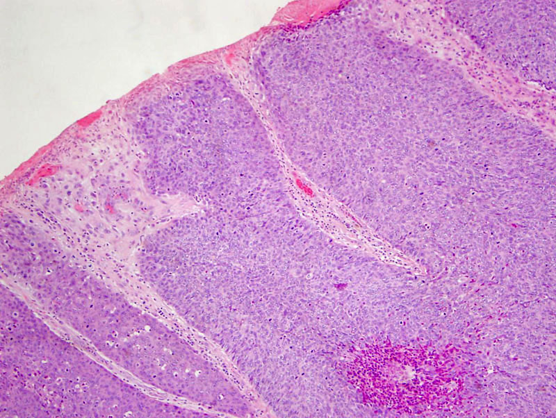
Case 2: This different case again demonstrates a proliferation of basaloid poroid cells.
Note the relationship to surface epithelium (top center) image. Dilated cystic structures and small sweat ducts can be helpful in making the diagnosis.
Nuclear pleomorphism, hyperchromasia and prominent nucleoli are features of malignant transformation.
Squamous metaplasia can be well appreciated here.
This is the malignant counterpart to a poroma. Also known as porocarcinomas, these tumors usually arise from benign poroid tumors and similar to poromas, are composed of neoplastic basaloid "poroid" cells.
In porocarcinoma, there is more pronounced cytologic atypia, hyperchromasia, mitotic activity, necrosis and an infiltrative growth pattern. Squamous metaplasia is a common feature (Busam, Obaidat). Not surprisingly, it may be difficult to distinguish a porocarcinoma with squamous metaplasia from a squamous cell carcinoma. Thus, one might consider highlighting the sweat duct epithelium with Cam5.2, CK7, Br-2 or CEA (Busam). Also, the presence of an adjacent benign poroma should be extremely helpful (if present).
These tumors carry a high risk of metastasis to the lymph nodes, lungs and other sites in the skin (Busam).
Busam KJ. Dermatopathology: Foundations in Diagnostic Pathology 1st Ed. Philadelphia, PA: Elsevier; 2010: 414-5.
Obaidat NA, et al. Skin adnexal neoplasms - part 2: An approach to tumors of cutaneous sweat glands. J Clin Pathol 2007;60:145-9.
)
)
)
.jpg', '', 'micro', ''))
)
)