System: Skin: Dermis: Neoplastic: Cutaneous Meningioma

System: Skin: Dermis: Neoplastic: Cutaneous Meningioma

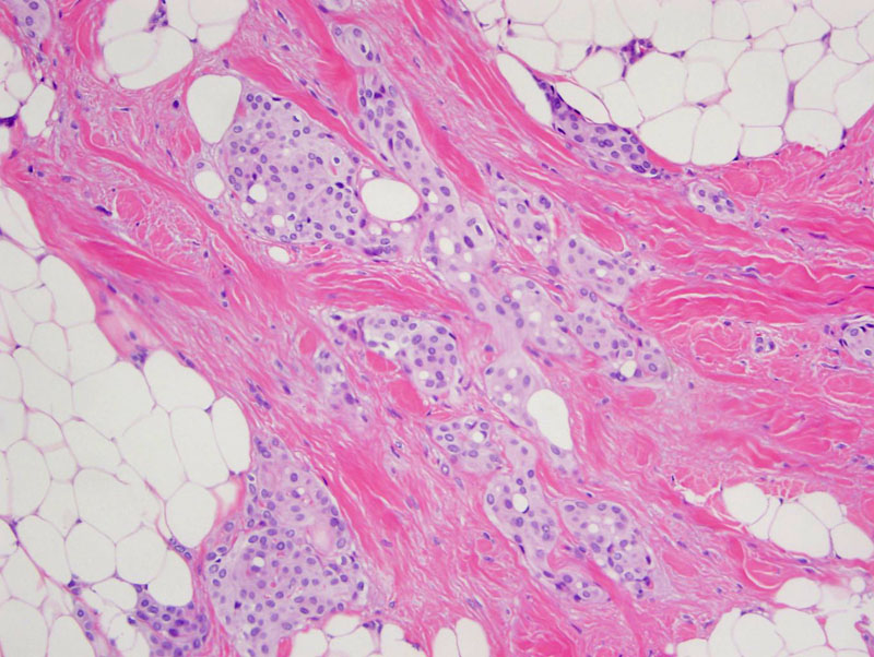
irregular clusters of uniform round cells with pink cytoplasm within the dermis Image
intranuclear inclusions in the meningioma; benign dermal structures too Image
Cutaneous meningioma is a rare neoplasm of the skin that is best classified into three types using the classification system proposed by Lopez et al in 1974.
Type I lesions are congenital and are most commonly present on the scalp, forehead, and paravertebral region at birth. They are thought to be derived from ectopic arachnoid cells that become trapped during fetal development. The terms ‘rudimentary meningocele’ and ‘acroleic meningocele’ have been used to describe type I cutaneous meningiomas.
Type II lesions most frequently occur on the face around the eyes, nose, and mouth as well as in the distribution of the cranial and spinal nerves. Given their cutaneous distribution, it is speculated that these lesions are derived from arachnoid cells which are deposited along the course of nerves after they penetrate the dura.
Type III lesions are the result of direct spread or distant cutaneous metastasis of a primary intracranial meningioma.
Cutaneous meningiomas typically present as skin colored, firm, subcutaneous nodules. As mentioned above, type I tumors are present at birth at are most commonly located on the scalp, forehead, and midline along the spine. Type II and III tumors present later in life, with type II lesions occurring around sensory organs of the head and in the distribution of cranial and spinal nerves, and type III lesions occurring on the scalp.
The majority of lesions are treated by surgical excision. It is important to remember that some lesions may be connected to the intracranial cavity, which may necessitate radiographic imaging prior to treatment.
Surgical excision with a clear margins typically curative for type I lesions. The prognosis for type III lesions is obviously worse as these result from local extension or distant metastasis of an intracranial meningioma.
Lopez DA, Silvers DN, Helwig EB. Cutaneous meningioma—a clinicopathologic study. Cancer. 1974;34:728–744.
Miedema JR, Zedek D. Cutaneous Meningioma. Arch Pathol Lab Med. 2012;136:208-11.
)
)