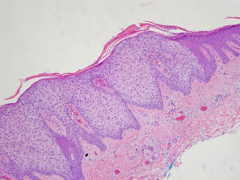

Case 1: The pale-staining keratinocytes are sharply demarcated from the non-lesional tissue. A psoriasiform proliferation with broad-based rete ridges is very common. The cells are slightly enlarged compared to their neighboring counterparts.
Note the presence of neutrophils in the epidermis. Neutrophilic abscesses, while not seen here, may be found in some lesions. A crusted scale on the surface of the epidermis is also very common.
Case 2: The pale keratinocytes are filled with glycogen.
Case 3: Transition from normal (right) to lesional pale-staining keratinocytes is demonstrated. The presence of neutrophils is characteristic, including collections neutrophils in the parakeratotic stratum corneum.
A closer look at the clear cytoplasm, inflammation. The pale keratinocytes are bland and rather uniform, as this is a benign neoplasm.
Clear cell acanthoma, also known as pale cell acanthoma, is a raised lesion consisting of pale-staining keratinocytes, most often in a psoriasiform growth pattern. The rete ridges are broad and somewhat centripetal. The lesion is sharply demarcated from normal adjacent epidermis with darker (normal) staining keratinocytes. The pale keratinocytes are glycogenated. Other features commonly seen include parakeratosis and neutrophils in the epidermis. Microabscesses may also form in the stratum corneum. Dilated blood vessels in the edematous dermal papillae may result in a red clinical appearance (Fletcher, Rapini).
Asymptomatic and seen as a red or flesh-colored dome-shaped papule. Arises in middle-aged to elderly adults and the most common location is the leg (Busam).
It is considered by "lumpers" to be an irritated (clear cell variant) form of seborrheic keratosis (Rapini), and clinically, it also has a "stuck-on" appearance.
Simple excision is curative.
→Key histologic features include pale keratincytes with expanded cytoplasm clearly demarcated from normal epidermis, acanthosis and exocystosis of neutrophils.
→Lesional cells lack phosphorylase, an enzyme necessary for the degradation of glycogen
Busam KJ. Dermatopathology: Foundations in Diagnostic Pathology 1st Ed. Philadelphia, PA: Elsevier; 2010: 344.
Fletcher CDM, ed. Diagnostic Histopathology of Tumors. 3rd Ed. Philadelphia, PA: Elsevier; 2007: 1427-8.
Rapini RP.Practical Dermatopathology. Philadelphia, PA: Elsevier; 2005: 238.