System: Gastrointestinal: Stomach: Autoimmune: Atrophic Gastritis

System: Gastrointestinal: Stomach: Autoimmune: Atrophic Gastritis

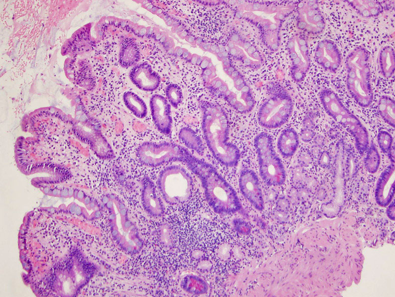
This patient has pernicious anemia resulting in this altered morphology in the gastric mucosa. Abnormal folds are found with a decrease in glands. The brightly eosinophilic neuroendocrine cells can be seen at the base. Intestinal metaplasia is also found.
Hyperplasia of the endocrine cells is easily appreciated.
Chromogranin highlights the endocrine cell population. The endocrine cells appear increased in number but fail to form a discrete mass.
Case 2 is a biopsy supposedly from the gastric fundus. The stain for gastrin was negative, confirming that the tissue is not antral in origin. Not that there are some small nests of cells deeper down in the lamina propria. Image
These cells stain strongly for chromogranin consistent with autoimmune atrophic gastritis. Image
)
)
)
)
)