
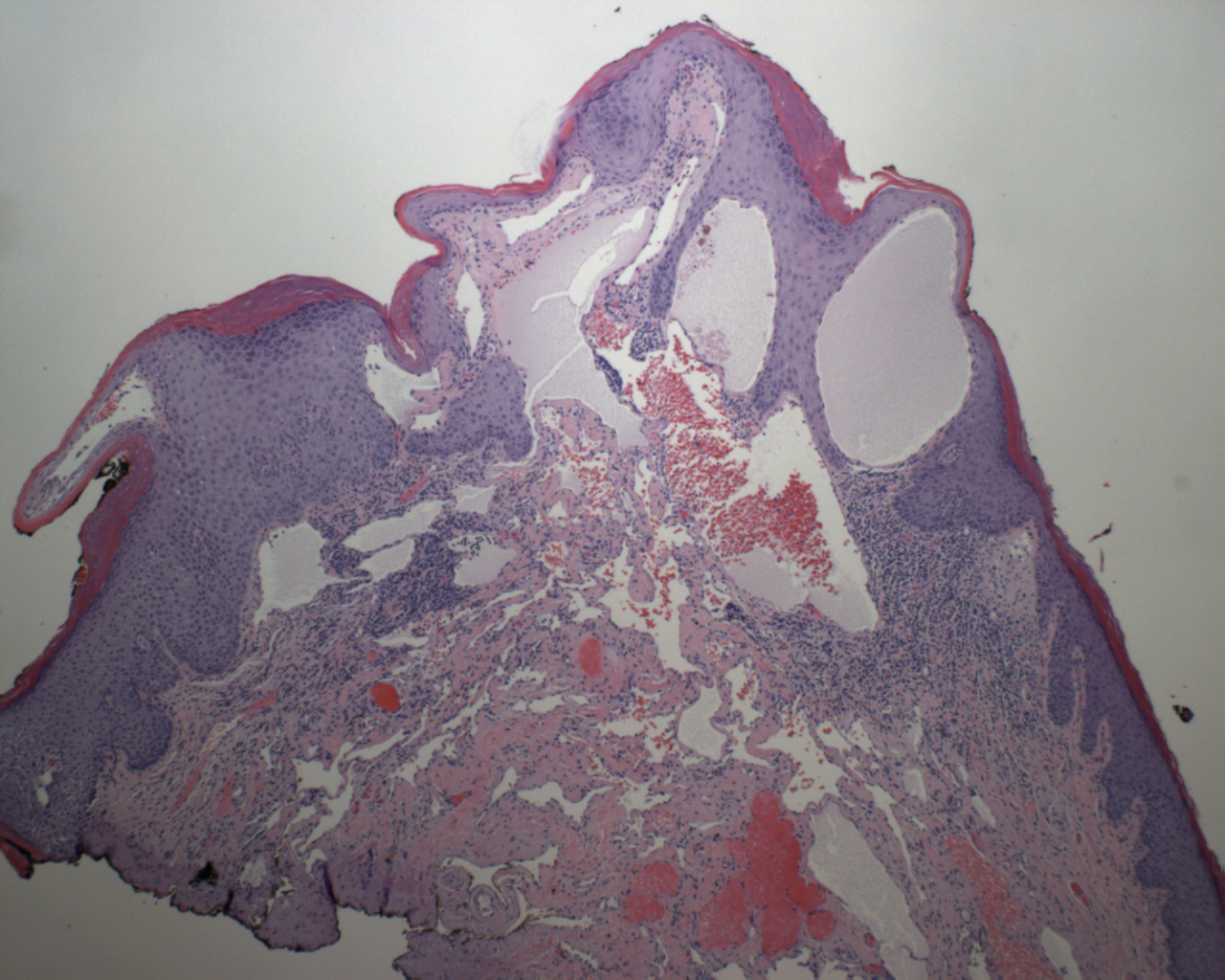
Dilated vessels appear concentrated in the papillary dermis with overlying keratosis.
)
Markedly dilated vessels are back-to-back and lined by bland endothelial cells.
)
The ectatic vessels form cavernous spaces, some of which may be filled with blood.
)
Angiokeratomas present clinically as small dark-red or purple raised spots. Histologically, they are characterized by ectatic dermal vessels with overlying irregularly acanthotic epidermis. Sometimes, the elongated rete ridges appear to embrace the ectatic vessels.
There are multiple subtypes of angiokeratomas, briefly:
(1) Sporadic angiokeratomas - common in middle age adults.
(2) Angiokeratoma circumscriptum - may be seen at birth as a "birthmark", but can appear in childhood or later. Clusters are seen in the trunk or extremities.
(3) Fordyce (scrotal) type - most common in elderly men, but a vulvar counterpart may be seen in younger women.
(4) Angiokeratoma corporis diffusum associated with Fabry syndrome - Fabry syndrome is an X-linked recessive lysosomal disorder. Patients have a deficiency of alpha-galatcosidase A leading to accumulation of ceramide trihexidose in lysosomes. In these patients, multiple angiokeratomas are seen in a groin and lower trunk.
In many instances, the lesions are present at birth, but they may first be seen in childhood or adulthood. The clinical impression shows telangiectasia, acanthosis, and hyperkeratosis. Sometimes they darken and clinically simulate melanoma.