System: Breast: Ductal: Neoplastic: DCIS, Cancerization of Lobules

System: Breast: Ductal: Neoplastic: DCIS, Cancerization of Lobules

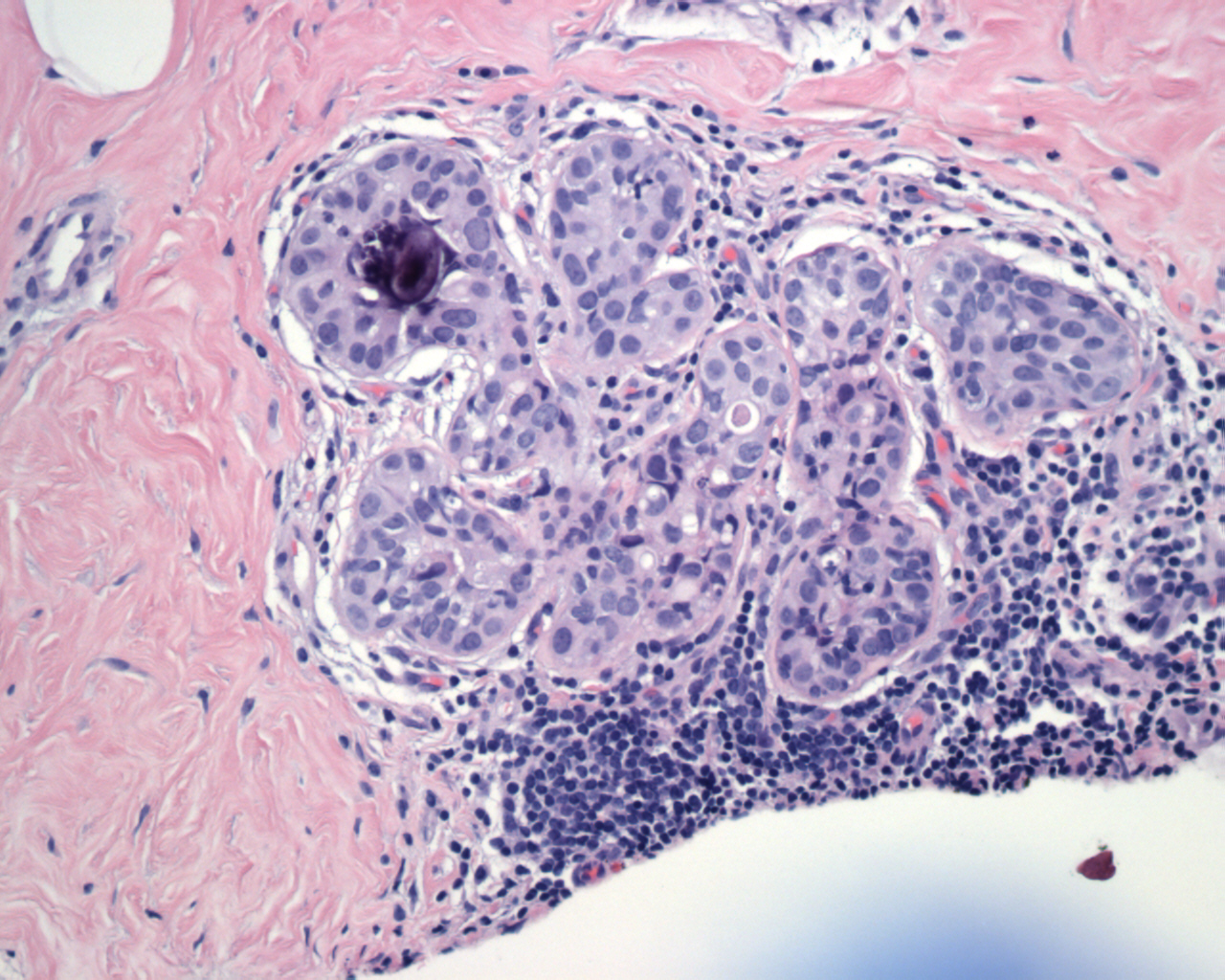
The edge of this breast section demonstrates cancerization of lobules. The cells are identical to the ones found in adjacent DCIS.
This is a duct involved by DCIS from the same case. The ductal structure is evident here. In many images of DCIS, the duct is simply a large circular structure, but it is actually a tubular structure.
At higher power, you can see that the morphology of the DCIS cells are identical to the morphology of those involving the lobules.
I had always been confused by this term. What it means, is that the neoplastic ductal cells have crawled backwards, so to speak, into the lobules. How to distinguish between LCIS and cancerization of lobules by DCIS? Compare the adjacent DCIS sure to be found in cancerization of lobules. The cell morphology should be similar to those in the ducts. The lobules will be involved by these cells, which are much more pleomorphic that the neoplastic cells seen in LCIS.
)
)
)