System: Gastrointestinal: Small Intestines: Reactive: Intestinal Lymphangiectasia

System: Gastrointestinal: Small Intestines: Reactive: Intestinal Lymphangiectasia

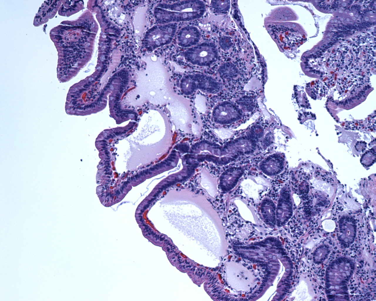
Low power view of a duodenal biopsy showing dilated lacteals. The overlying surface epithelium is normal.
Dilated lymphatics can be located in the mucosa or submucosa.
Intestinal lymphangiectasia is characterized by dilated intestinal lacteals. Most cases have been described in young children in association with other congenital abnormalities, but this finding is increasingly noted in older adults during endoscopy. Intestinal lymphangiectasia can be primary or secondary, with the latter being much more common in adults.
Thus, in adults, a variety of conditions associated with lymphangiectasia must be investigated. This includes a malignancy (e.g. lymphoma) which may obstruct the lymphatic system, infectious (Whipple's disease) and inflammatory bowel disease (Crohn’s disease). The dilated lacteals result in loss of lymph fluide into the bowel lumen, leading to hypoproteinemia, hypogammaglobulinemia, hypoalbuminemia and lymphopenia.
Histologically, dilated lymphatics involve the mucosa and submucosa. The dilated lacteals may be focal.
Clinical findings include bilateral lower limb edema, pleural effusion, chylous ascites and iron deficiency anemia (Freeman). In fact, the patient from this has had longstanding iron deficiency anemia. The cause of anemia in intestinal lymphangiectasia is not entirely clear and may be related to malabsorption of iron or angiodysplasia (Freeman).
A low fat diet and supplementation with medium-chain triglycerides, which bypass the intestinal lacetal system.
Freeman HJ, Nimmo M. Intestinal lymphangiectasia in adults. World J Gastrointest Oncol. 2011 Feb 15;3(2):19-23.
)
)