System: Genitourinary: Testis: Neoplastic: Sertoli Cell Tumor

System: Genitourinary: Testis: Neoplastic: Sertoli Cell Tumor

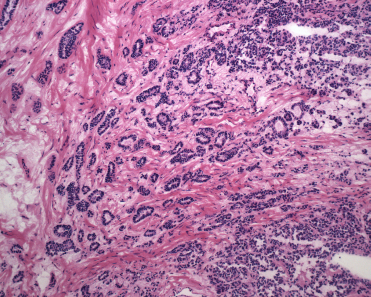
The neoplastic Sertoli cells form tubules.
Looser, more edematous areas are admixed with more cellular regions.
The tubules may form glandular structures or may be compressed to form more solid areas.
The neoplastic tubules had focal positivity for cytokeratin and was negative for PLAP and OCT4.
Sex-cord stromal tumors in the testis are rare and comprise ~5% of testicular tumors (Humphrey). Members of this group include Leydig cell tumors, Sertoli cell tumor, granulosa cell tumor, thecoma and fibromas.
Sertoli cells comprise ~1% of all testicular tumors. Grossly, the mass is well-circumscribed with tan-yellow hemorrhagic cut surface. Histologically, bland cells are most often arranged in tubules with occasionally retiform (net-like), tubular-glandular and solid patterns.
Subtypes include lipid rich, large cell calcifying and scleorsing variants.
In terms of IHC, sertoli cell tumors are usually cytokeratin and occasionally inhibin positive. They are negative for PLAP and OCT4.
• Testis : Large Cell Calcifying Sertoli Tumor
)
)
)
)